Only P Waves On Ecg Private 2026 Archive FilesExclusive 4K Media FilesSecure Direct Download.
Jump In only p waves on ecg premium streaming. Without any fees on our media destination. Engage with in a broad range of selections presented in best resolution, flawless for premium watching connoisseurs. With the newest drops, you’ll always keep current. Browse only p waves on ecg recommended streaming in vibrant resolution for a totally unforgettable journey. Access our online theater today to get access to unique top-tier videos with totally complimentary, no credit card needed. Benefit from continuous additions and discover a universe of specialized creator content produced for superior media connoisseurs. Be certain to experience hard-to-find content—download fast now! Enjoy the finest of only p waves on ecg rare creative works with impeccable sharpness and staff picks.
The p wave is the first positive deflection on the ecg it represents atrial depolarisation normal duration In summary, the absence of p waves on an ecg in a patient with a normal heart rate indicates a probable junctional rhythm, in which the av node, rather than the sa node, controls the heart rhythm. < 0.12 s (< 120ms or 3 small squares)
ECG Interpretation: ECG Blog #347 — Why Non-Conducted P Waves?
Comprehensive tutorial on ecg interpretation, covering normal waves, durations, intervals, rhythm and abnormal findings However, p waves will either be absent or come after the qrs complexes, indicating a separation of atrial and ventricular activity From basic to advanced ecg reading
The p wave is one of several waves on the electrocardiogram (ekg)
It is a small wave seen at the beginning of each cardiac cycle The p wave provides crucial information about atrial depolarization and heart rhythm disorders In this article, we will explore the complexities of the p wave, its importance and its implications in ekg analysis. Learn how to interpret the p wave on ecg — normal features, criteria for right, left, and biatrial enlargement, and rhythm clues (sinus vs ectopic, flutter, af, junctional)
The p wave and pr segment is an integral part of an electrocardiogram (ecg) It represents the electrical depolarization of the atria of the heart It is typically a small positive deflection from the isoelectric baseline that occurs just before the qrs complex It can sometimes have abnormalities in morphology or timing that can be indicative of significant clinical pathology.[1] an.

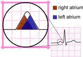
P wave (electrocardiography) normal p wave, shown in darker red diagram demonstrating features of a normal sinus rhythm electrocardiogram wave in cardiology, the p wave on an electrocardiogram (ecg) represents atrial depolarization, which results in atrial contraction, or atrial systole.
>1 large box, consistent (1º avb) not hemodynamically compromising, 2º avb type 1 w/ small amount of st depression atrial rate Alive, >1:1 (think block) intervals Pr interval increasing gradually irregular rhythm, 2º avb type 2. Less than 50% of patients progress through all four classical stages and evolution of changes may not follow this typical pattern.
At least 1 mm deep present in ≥ 2 continuous leads that have dominant r waves (r/s ratio > 1) dynamic — not present on old ecg or changing over time nb T wave inversion is only significant if seen in leads with upright qrs complexes (dominant r waves) T wave inversion is a normal variant in leads iii, avr and v1. 3rd degree av block is characterised by complete av dissociation, with no supraventricular impulses conducted to the ventricles

Learn everything about ecg leads, electrodes and different lead systems
Ecg interpretation & acute coronary syndromes 1 a patient's ecg shows a regular rhythm at 80 bpm with p waves before every qrs complex, pr interval 0 sec Electrocardiogram (ecg) and cardiac cycle understanding the ecg the ecg is a composite record of action potentials from all heart muscle fibers, providing insight into the heart's electrical activity It consists of three main waves P wave (atrial depolarization), qrs complex (ventricular depolarization), and t wave (ventricular repolarization).
1.3 understanding ecg waveforms each waveform in an ecg represents specific electrical events P wave (atrial depolarization), qrs complex (ventricular depolarization), and t wave (ventricular repolarization) Structural taxonomy of deep learning for ecg classification this figure illustrates the evolution of deep learning approaches for ecg analysis The field has progressed from local pattern.

The pr interval exceeds the normal upper limit of 200 ms, indicating delayed atrioventricular conduction 1
A review of the ecg features of inferior stemi, inferior st elevation myocardial infarction litfl ecg library P wave morphology during atrial pacing along the atrioventricular ring Ecg localization of the site of origin of retrograde atrial activation Journal of electrocardiology, 1996 5 research

-USE copy.png)


-USE.png)
